Датотека:Human brain anterior-inferior view description.JPG
Human_brain_anterior-inferior_view_description.JPG (330 × 475 пиксела, величина датотеке: 31 kB, MIME тип: image/jpeg)
| Ово је датотека са Викимедијине оставе. Информације са њене странице са описом приказане су испод. Викимедијина остава је складиште слободно лиценциралних мултимедијалних датотека. И Ви можете да помогнете. |
Опис измене
| Опис |
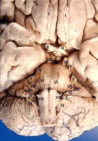
Human brain - anterior-inferior view - cerebral nerves I. N. olfactorius - The fila olfactoria (approximately 20 nerve rootlets on each side) comprise the Olfactory nerves (I) which run from the olfactory receptors in the nasal cavity to the olfactory bulb. II. N. opticus - The Optic nerves (II) are large & join at the midline to form the optic chiasm, then continue laterally as the optic tracts. The optic nerve is not a true nerve but rather a CNS tract. III. N. oculomotorius - The Oculomotor nerves (III) emerge from a depression in the midbrain, the interpeduncular fossa, just caudal to the optic chiasm. IV. N. trochlearis - The Trochlear nerves (IV) are small & are seen on the lateral surface of the midbrain. They are the only nerves which exit from the posterior side of the brain. V. N. trigeminus - The Trigeminal nerves (V) are large and emanate from the lateral surface of the pons. VI. N. abducens - The Abducens nerves (VI) exit near the midline from the inferior pontine sulcus which separates the pons from the medulla. VII. N. facialis - Moving laterally in the inferior pontine sulcus, the Facial nerves (VII) can be seen. VIII. N. vestibulocochlearis - Slightly lateral to the facial nerve is the Vestibulocochlear nerve (VIII). IX. N. glossopharyngeus - The postolivary sulcus is a groove running rostrocaudally on the lateral surface of the medulla. From this sulcus pass the small Glossopharyngeal nerves (IX) rostrally and... X. N. vagus - ...the much larger Vagus nerves (X) caudally. XI. N. accessorius - The Spinal Accessory nerves (XI) exit the cervical cord then pass rostrally through the foramen magnum to exit the cranial vault with the lossopharyngeal and vagus nerves. XII. N. hypoglossus - The Hypoglossal nerves (XII) exit the medulla via the preolivary sulcus.
|
| Датум | |
| Извор | http://www.healcentral.org/healapp/showMetadata?metadataId=40566 (Internet Archive of file description page) |
| Аутор |
John A Beal, PhD Dep't. of Cellular Biology & Anatomy, Louisiana State University Health Sciences Center Shreveport |
| Дозвола (Поновно коришћење ове датотеке) |
CC-BY |
| Остале верзије |
 |
Лиценцирање
- Дозвољено је:
- да делите – да умножавате, расподељујете и преносите дело
- да прерађујете – да прерадите дело
- Под следећим условима:
- ауторство – Морате да дате одговарајуће заслуге, обезбедите везу ка лиценци и назначите да ли су измене направљене. Можете то урадити на било који разуман манир, али не на начин који предлаже да лиценцатор одобрава вас или ваше коришћење.
This file, which was originally posted to
http://www.healcentral.org/healapp/showMetadata?metadataId=40566, was reviewed on 25 September 2013 by reviewer Eleassar, who confirmed that it was available there under the stated license on that date.
|
Историја датотеке
Кликните на датум/време да бисте видели тадашњу верзију датотеке.
| Датум/време | Минијатура | Димензије | Корисник | Коментар | |
|---|---|---|---|---|---|
| тренутна | 20:22, 24. јун 2006. | | 330 × 475 (31 kB) | Patho | {{Information| |Description='''Human brain - anterior-inferior view - cerebral nerves''' I. N. olfactorius - The fila olfactoria (approximately 20 nerve rootlets on each side) comprise the Olfactory nerves (I) which run from the olfactory receptors in |
Употреба датотеке
Следећа страница користи ову датотеку:
Глобална употреба датотеке
Други викији који користе ову датотеку:
- Употреба на ar.wikipedia.org
- Употреба на azb.wikipedia.org
- Употреба на az.wikipedia.org
- Употреба на bs.wikipedia.org
- Употреба на de.wikipedia.org
- Употреба на de.wikibooks.org
- Употреба на en.wikipedia.org
- Cranial nerves
- User talk:Hovea
- User talk:Wouterstomp
- User talk:Nephron
- User talk:NCurse
- User talk:Robotsintrouble
- User talk:Was a bee
- User talk:Bloomingdedalus
- User talk:Read-write-services
- User talk:Attys
- User talk:Bakerstmd
- User talk:Manfi
- User talk:Mikepascoe
- User talk:Vokesk
- User talk:Neuraxıs
- User talk:Cmungall
- User talk:Qxukhgiels
- User talk:DocElisa
- User talk:JakobSteenberg/Archives/1
- User talk:Meteor sandwich yum
- User talk:Slashedone
- User talk:Anindya07
- User talk:Mattimussi
- User talk:Jelly Bean MD
- User talk:Benrusholme
- User talk:Brad.w.english
- User talk:Krsna ss
- User talk:Tom (LT)/Archive 4
- User talk:Dregon131
- User talk:Sapientia42
- User talk:Tilifa Ocaufa
- User talk:Snow Rise/Archive 6
- User talk:Arcadian
- User talk:Outofbattery
- User talk:Anatomyczar
- Wikipedia:WikiProject Anatomy/Newsletter/4
- User talk:Supravibhatsupravi
- User talk:Athikhun.suw
- User talk:CFCF/Archive 6
- User talk:Lewisskinner/ArchiveJul 2015
- User talk:Tyrol5/Archive 6
- User talk:CFCF/Archive 7
- User talk:DiverDave/Archive 5
Још глобалног коришћења ове датотеке.